
I want to see what is happening after conceiving inside my baby bump? Will I do sonography so early? Taking the ultrasound before has created many questions in your subconscious mind.
“what does an ultrasound at 6w5d look like?” Be relax. The content is arranged for you.
The ultrasonography process helps verify the embryo condition and 1st day of the last period. Doctors suggest a scan due to irregular menstruation or any physical complication.
Before getting an ultrasound, you should consider a few more things that will help keep you stress-free. So let’s dive in!
What does an ultrasound at 6w5d look like?
Initially, it was a little hesitant about what was held in the abdomen. After emptying the bladder, it is seen through the ultrasound of your stomach. You got the bed so that it can be easy to see on the screen that it is going to be like an unpainted something.
First picture of ultrasonography
The first ultrasound images can be seen in a digital way, but the color is rarely expressed. So you can collect the picture with the permission of the technicians in that case.
The scans system is used to print the whole facts pictures in the black and white mood in the form of reports. In this way, you get the correct facts that can be understood according to the image.
Sonography procedure
Transvaginal ultrasound performed is usually done vaginally with the help of the trimester. The doctor inserts a tampon-length transducer that is comfortable. Then check around the abdominal area. The ultrasound tests are done from 0 to 22 weeks
Overall, the whole thing doesn’t take more than 4-5 minutes. Technologists can verify the fetal pole condition. But you can’t match it on the screen in that way.
Although it may assume a few more weeks to see precisely the embryo to appear, you can see the empty pouch next to the fetus’ heartbeat. It’s not bad in the early time.
The sonographer will confirm the condition of the fetus and the arrival date of the baby. Besides, check the length of the sample, uterus, or the fallopian tube. It will examine when your tubes are tied but you have late period. It may be 2-3 days, which can be plus or minus in this case.
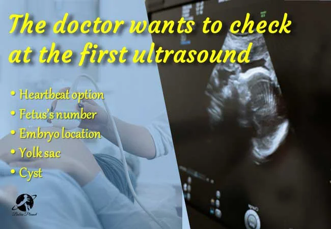
The doctor wants to check at the first ultrasound
The fetus situation and some essential issues stem from selecting the upcoming baby.
6w5d ultrasound heartbeat
The sonographers try to see the pulsation on the screen. Mainly, six weeks are suitable for feeling the heart form. It is an emotional segment for the first time of pregnancy.
Fetus’s number
A fetus may be assured to have twins or more embryos instead. The twin result comes up at 4-5 weeks of pregnancy. The rare case is this story is complete, so this side is noticed. However, it is easier to understand after checking it. It may take a few more days to know in some cases.
Embryo location
The embryo has been implanted in the uterus that is being verified. It is examined to see if it is possible to be placed outside the uterus. Or in the fallopian tube.
Thus, it can lead to ectopic pregnancy or other complications. So this is important to be sure.
Yolk sac
The sonographer can see a yolk sac inside the gestational sac. The sac looks like a tiny balloon that includes the fetus. Some times the Yolk sac looks empty too.
The doctor checks its shape and health condition through this. It is also merged into a human child at the end of 12 weeks.
Cyst
The technician examines the corpus luteum cyst, and it usually stays. The cyst is formed on the follicles, the egg release site.
The scanning process is used to diagnose the condition and observe the sign of the sac.
What if no baby is visible at the ultrasound?
Primarily can find no heartbeat in 6 weeks through ultrasound. The challenging issue is possible when ovulation is not going on properly. It can be seen in 3-6 weeks on the transvaginal ultrasound, but some things may differ.
Your date calculation may be incorrect or irregular menstruation, making it invisible. Now, may calculate another 1 -2 weeks from your healthcare provider for the next ultrasound. Most abortions are possible within six weeks of pregnancy.
By the way, there are some exception when you may not see the fetal pole but still you will have the success stories. For example, it may happen at 6 weeks or even 7 weeks.
The option may be the last menstrual period that is beyond your expectations. However, once the fetal pulse is ensured, it is the key to avoiding miscarriage.
The fetal is so tiny that there is no option to see or feel it with the naked eye in some cases. It can be a quarter of an inch long, which is tough to see in the first stage. However, it can be seen after a print snapshot that can be visible.
Are ultrasounds should this early?
The month or week of pregnancy date of birth is determined by ultrasound. However, it has an estimated time. It can help to ensure the health and well-being of the fetus.
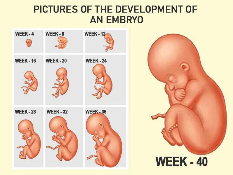
In the initial stage, it did not determine the cause of the ultrasound damage as far as yet is concerned. It can take pictures of the development of an embryo child using sound waves without any harmful radiation.
“The time of ultrasound is done can raise the temperature by 4°C degrees Celsius” claimed in an journal published in National Library in Medicine . You get some tremendous excessive points, so there is no way to complain about it.
However, it gives good feedback to keep the month count right of alertness on the mom.
When should ultrasound be done during pregnancy?
If the period is regular and the date is known, then early ultrasound is not suggested. It is usually preferred for 14 weeks or three months to learn a clear idea from the ultrasound. However, this may not be common in all areas.
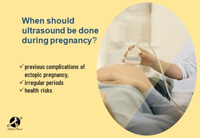
So when should ultrasound be done during pregnancy? Early ultrasound is essential when you have any previous complications of ectopic pregnancy, irregular periods, or health risks. Otherwise, there may be a tension grooming of pain, miscarriage, & bleeding.
The doctor fixes it by physically examining the body or condition. It brings excitement in the case of new pregnancies or sickness. The question remains, what can happen?
The correct calculation period is determined from the first menstrual period to pregnancy time and continuum.
What can ultrasound show in 8 weeks?
This is the first time to detect the onset of embryo pulsation in 6 weeks. The sonography should first be visible to an embryo and throb according to the counting date. You cannot feel the show-off in some cases that are not different.
The weight, blood pressure, blood type and fitness, physical condition are tested in this case. It’s right to see a natural human feature that is emotional. Then check if there is any abnormality in the baby.
What can be seen in the ultrasound of 1 week?
Basically, the pregnancy tested result is located at the initial stage. You have also contained a blastocyst not seen in a developing placenta. Also, there is no pulsation and nothing to see.
The process can see small scans in the field of scans that are not easily visible. It can confirm pregnancy with transvaginal ultrasound. However, it is also a useful tool for giving a quick pride check for specific results.
When can a heartbeat be appropriately seen?
The ultrasound in female private part essentially aids in the detection of embryo. But the time of 5-6 weeks run, that time is determined to view the fetus. It can look like a sac or visible sign.
The fertilized egg development & pulse are diagnosed easily in that time. Thus, the fetal pole success story comes out at 3 to 8 weeks of pregnancy (Embryo development timeline).
To Conclude
The ultrasound of 6 weeks 5d is as essential in the pregnancy stage as coming on excitement. There is an option to count the months until the baby’s birth carefully.
You will see something visible that is going to indicate a human fetus. The doctor may not be allowed to say all things to give a scan picture to you.
It is better to look at the things you need without hesitation in that case. However, the health providers are expressing essential things with giving direction for stress-free. Happy pregnancy journey.
Source:
Kurban Y, Uyar I, Alan M, Hacifazlioglu C. Fetal sex prediction measuring yolk sac size and yolk sac-fetal pole distance in the first trimester via ultrasound screening. J Ultrasound. 2021 Dec;24(4):489-492. doi: 10.1007/s40477-020-00516-0. Epub 2020 Nov 25. PMID: 33237452; PMCID: PMC8572229.